Read more at: Alumni stories: Greg Reeves
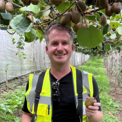
Alumni stories: Greg Reeves
10 June 2025
Studying Plant Sciences at Cambridge can open up a wealth of career opportunities. In this series, we hear from Plant Sciences alumni about their experiences of studying here, how it shaped their careers and what they are doing now. If you would like your alumni story featured, please email us at alumni@plantsci.cam.ac.uk...